Realignment osteotomy, microfracture and chondroplasty
Knee alignment has a profound effect on the forces going through each compartment of the knee. Varus alignment (bow legs) increases the forces on the medial (inside) half of the knee and valgus alignment (knock knees) increases the forces of the lateral (outside) half of the knee. With progressive arthritis is an isolated compartment, the malalignment worsens, which further exacerbates the arthritic symptoms.
 In younger (<60yo), active individuals with isolated arthritis and associated malalignment (usually medial arthritis and bow leg), correction (or slight overcorrection) of the alignment can unload the arthritic compartment and take the weight through the non-arthritic part of the knee. This aims to provide symptomatic relief whilst preserving the native knee and delaying the need for knee replacement in the medium future.
The osteotomy (cut bone) is usually done in the proximal tibia (shin bone), so is known as a high tibial osteotomy (HTO). It involves partially cutting the bone under controlled conditions, correcting the alignment and then fixing the newly created “fracture” with a plate and screws until the osteotomy heals.
Osteotomy is usually combined with simultaneous arthroscopy to assess the wear and tear in the remainder of the knee and treat any associated mechanical symptoms that arise from meniscal tears or loose bodies. Localised cartilage damage can be managed with microfracture (drilling the base of the cartilage defect to stimulate the underlying bone marrow healing response) or chondroplasty (trimming and sealing the edges of localised cartilage damage to attempt to prevent propagation of loose cartilage flaps).


 Other knee ligaments include the posterior cruciate ligament (PCL), medial collateral ligament (MCL) and posterolateral corner (PLC) complex made up of the lateral collateral ligament (LCL) and popliteus tendon. Finally, the quadriceps tendon (thigh muscle into knee cap) and patella tendon (knee cap into shin bone) complete the extensor mechanism attachments around the knee.
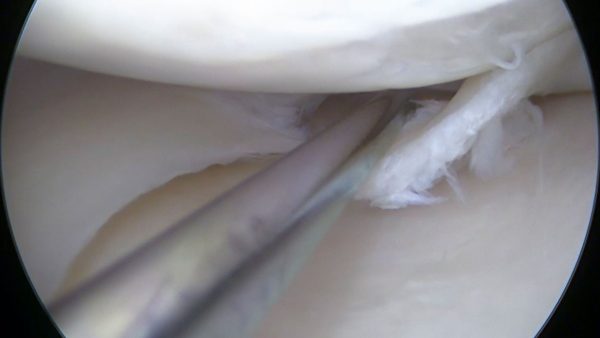 Meniscal tears in younger people (<40yo) are often the result of acute trauma in an otherwise normal meniscus. Early repair, if successful, allows the meniscus to return to its normal functions. In older people, or chronic tears, the meniscus is usually sufficiently damaged that repair is less likely to be successful. In these cases, excision of the torn fragments can improve mechanical and synovitis symptoms from the tear or associated meniscal cysts. If the knee already has significant arthritis symptoms, then meniscal surgery is rarely indicated.
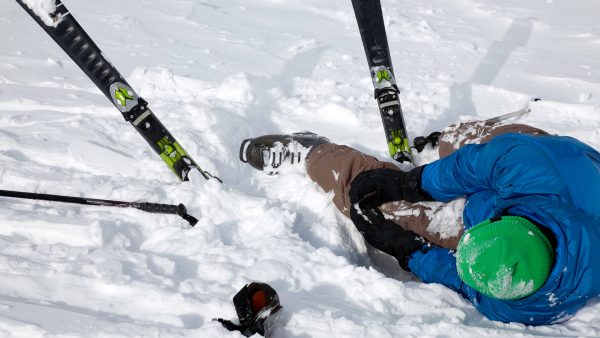 Acute knee injuries range from minor sprains and swelling (effusions) to complete knee dislocations which can compromise the nerve and blood supply to the entire leg. As such, a detailed history of the injury and preinjury function, along with a focused clinical examination and investigations (Xray and/or MRI) is required before the diagnosis can be made and specific management recommended. In some cases, a specific brace and/or physiotherapy program may yield good functional outcomes, whilst others may require early surgical repair or reconstruction.
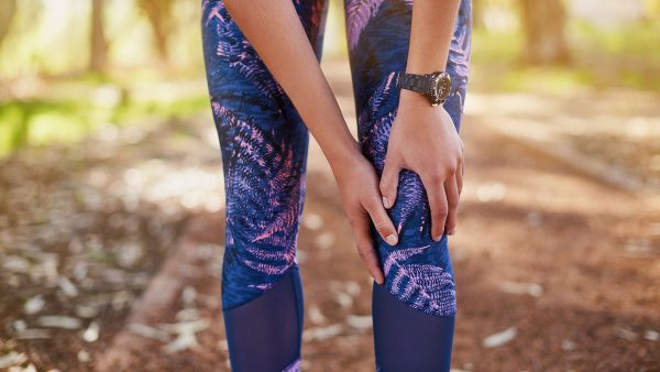 Recurrent instability is uncommon after adequate rehabilitation unless specific biomechanical parameters are present. These include patients with trochlea dysplasia (shallow groove), patella alta (high patella), lateralised tibial tubercle (increase Quadriceps “Q” angle) and ligamentous laxity (“double jointed” or high flexibility”). In these cases, persistent instability can be very debilitating, so patellofemoral stabilisation including realignment surgery to correct the above parameters (often tibial tubercle transfer), combined with MPFL reconstruction (using a hamstring tendon) is indicated.
 Osteoarthritis is the destructive process in which joint cartilage wears out, leaving the bones rubbing directly on each other. This is associated with variable degrees of pain and stiffness. Joint replacement (arthroplasty) surgery involves cutting away worn out bone and cartilage and replacing those articulating surfaces with a combination of metallic knee implants and highly specialised plastic (polyethylene).
 In some cases, the arthritis and pain are localised to only one compartment (most commonly medial) and so only that compartment needs to be replaced. This is called Unicompartmental (“uni” or “partial”) knee replacement. In properly selected patients this procedure is functionally superior to total knee replacement, has an easier recovery and a lower risk of major complications. However, it has the risk of requiring revision surgery to a total knee replacement and many patients are not suitable for specific reasons.